Table of Content
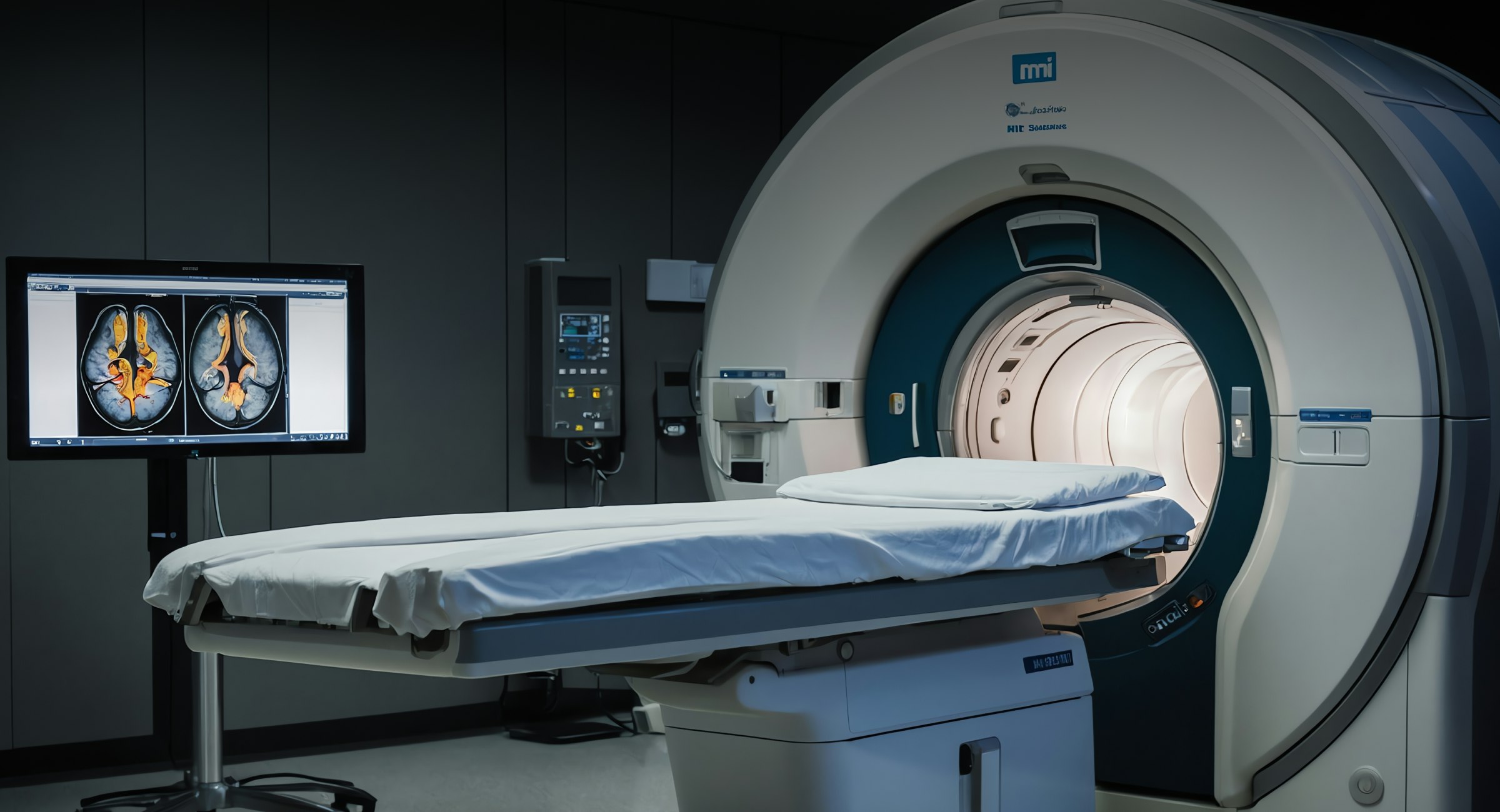
Medical imaging, like CT scans and MRIs, is a huge part of modern healthcare, involving medical images. It’s non-invasive, accurate, and beneficial for catching diseases early. It has made giving patients the best care possible easier.
But, even after years of training, the human eye can miss small details in these images, which could lead to mistakes in diagnosis.

This is where Machine Learning (ML) steps in.
It’s a powerful technology that can spot even the tiniest abnormalities and automate some of medicine’s more repetitive tasks like image processing.
Curious to see how it works in detail?
In this blog post, we’ll walk through some of the most popular ways ML is used in medical imaging and the benefits it brings to healthcare companies.
Machine Learning Development Services!
Explore Now!
Use Cases of Machine Learning (ML) in Medical Imaging Analysis
1. Cancer detection and diagnosis
ML is changing the game when it comes to cancer detection across various types of cancers. In prostate cancer, for instance, it’s used with MRI and biopsy images to pinpoint tumor locations and figure out how aggressive the disease might be.
For lung cancer, ML helps catch tiny nodules on chest X-rays or CT scans that a radiologist might miss. The technology is also great for skin cancer, where ML can analyze dermoscopic images to tell the difference between malignant spots and benign ones.
The power behind this?
Neural networks like Convolutional Neural Networks (CNNs).
Simply put: these networks are amazing at recognizing patterns in images.
Take breast cancer, for example—Deep learning algorithms like CNNs are trained using thousands of mammograms to spot suspicious masses.
They start by learning simple shapes and work their way up to detecting more complicated features, simplifying the process of finding potential tumors.
But ML algorithm development doesn’t stop here.
They can also analyze high-resolution tissue slides to distinguish between normal and cancerous cells. The result? Faster and more accurate diagnoses, giving healthcare providers a head start on pharma treatment decisions.
2. Brain tumor segmentation
In the early stages, brain tumors can look almost identical to healthy tissue, making it hard to spot them on Magnetic Resonance Imaging (MRI). Manual segmentation of these scans can take hours per patient, which is exhausting and leaves room for errors.
Plus, different radiologists might see the tumor in slightly different ways due to fatigue, experience, or just the trickiness of interpreting ambiguous areas in the scan. That variability can lead to inconsistent treatment plans.
This is where ML makes a difference.
U-Net, a deep learning model, is a massive help because it can work with fewer labeled images and still deliver accurate results. It combines high-level image features with spatial data to outline the boundaries of the tumor more clearly.
There are even deep learning models, like 3D U-Net and V-Net, which examine 3D MRI scans (instead of just 2D slices) and perform image synthesis to better understand the tumor’s shape and location across multiple layers of the brain.
Then you’ve got DeepMedic, another neural network model that processes MRI images at different scales, making it perfect for tumors of all shapes and sizes.
ML-assisted medical image segmentation improves surgical planning and treatment in a few key ways:
- Accurate tumor boundaries—critical for neurosurgeons when they’re planning surgeries
- Personalized radiation therapy, which involves targeting just the tumor while protecting nearby healthy tissue
- Better post-surgery care, including precision medicines and personalized adjustments to treatment after surgery
3. Cardiac imaging and disease prediction
ML has become a fantastic tool in cardiac imaging, simplifying processes that once required manual work from cardiologists.
Here’s how it helps:
ML algorithms, especially CNNs, can analyze echocardiogram images to automatically detect important features like left ventricular ejection fraction (LVEF), valve issues, and wall motion problems. This means more consistent assessments compared to traditional methods.
In cardiac CT scans, ML models identify coronary plaques, segment arteries, and measure stenosis, making the detection of coronary artery disease (CAD) much simpler.
These algorithms can also combine imaging data with clinical factors (like patient age or cholesterol levels) to calculate a patient’s risk for CAD. They can forecast disease progression and predict the likelihood of heart failure.
When it comes to cardiac MRI, ML provides a highly detailed view of the heart. It can recognize conditions like myocardial infarction, cardiomyopathy, or fibrosis and can segment heart chambers to measure size, volume, and myocardial thickness.
The benefit?
Faster, more reliable diagnoses and the ability to tailor treatments based on individual risk profiles in clinical settings.
With such extensive data at their fingertips, cardiologists can prioritize patients who need urgent care and initiate targeted treatment plans. This improves hospital efficiency, cuts down on patient expenses, and smooths workflows for everyone involved.
4. Retinal disease screening
ML is making a big impact in ophthalmology, helping detect and diagnose retinal diseases more quickly and accurately.
For starters, CNNs can analyze fundus images to spot abnormal retinal structures. They can identify signs of Diabetic Retinopathy (DR), like hemorrhages and microaneurysms, and even grade the severity of the disease automatically.
ML can analyze Optical Coherence Tomography (OCT) images by looking at the optic nerve head and the retinal nerve fiber layer (RNFL) thickness. This makes it easier to catch early signs of glaucoma.
Of course, ML models can work with OCT scans to recognize drusen (yellow deposits) or abnormal blood vessels, which are classic signs of age-related macular degeneration (AMD).
ML offers something huge here—scalability. It can process large volumes of retinal images quickly and accurately, making it perfect for large-scale screening programs, especially for high-risk groups like those with diabetes.
Beyond that, this advanced technology has the potential to bring retinal disease screening to rural or underserved areas with limited access to specialists.
These ML-powered programs can run in primary care offices, pharmacies, or even mobile clinics with very little human oversight. Portable cameras can capture retinal images, and ML algorithms handle the rest, analyzing the images and delivering results quickly.

5. Lung nodule detection
ML has proven helpful in detecting lung nodules, which are small, abnormal growths in the lung that signal early-stage cancer.
Using CNNs, ML models are highly sensitive to spotting nodules on chest X-rays. They’re trained to recognize patterns that may indicate the presence of nodules, even if they’re tiny or hidden behind other structures.
In regards to CT scans, 3D CNNs go even further by providing detailed information on the nodule’s shape, size, texture, and location. ML algorithms process these features to improve the accuracy of distinguishing between benign and cancerous growths.
They can even combine imaging data with clinical factors—such as age, smoking history, and genetic background—to give a clearer picture of which nodules need immediate attention.
This is a huge plus when screening large, high-risk populations, like smokers or those with a family history of lung cancer.
Another good news is that many healthcare providers are now integrating ML-powered Computer-Aided Diagnosis (CAD) systems into their radiology departments, providing a confidence score for each detected nodule. This means fewer false positives compared to manual interpretations alone.
6. Bone fracture detection
Imagine if there was technology that could spot fine yet critical fractures around joints, spines, and ribs that the human eye would otherwise miss.
ML models are trained on large datasets to automatically analyze X-rays and detect dislocations or hairline cracks with greater accuracy. This is especially useful in busy emergency departments, where time is critical and the healthcare providers are overworked or tired.
In fast-paced environments like trauma centers, ML systems can handle a high volume of patient data, allowing healthcare providers to offer help in situations like mass accidents, where immediate and accurate fracture detection can prevent delays in treatment.
And did you know that ML algorithms don’t just classify X-rays as “fractured” or “non-fractured?”
That’s right!
They also analyze detailed features like bone alignment and dislocations and even provide a breakdown of the fracture type (e.g., simple, compound, or displaced).
For hospitals without access to specialized radiologists, especially in rural areas, ML can serve as a valuable diagnostic aid for general practitioners or junior doctors who may not have extensive experience in interpreting complex cases.
The system can alert them to potential fractures and suggest further investigation or specialist referral.
7. Classification of neurological disorders
ML is making waves in neurology by automating the analysis of brain imaging data from MRI, fMRI, and PET scans. It’s helping detect and classify conditions like Alzheimer’s, Parkinson’s, and multiple sclerosis (MS) more quickly and accurately.
One of the biggest benefits?
ML can detect subtle brain changes long before clinical symptoms appear. This early detection means earlier interventions, which can slow the disease’s progression and improve patient outcomes via relevant drug discovery and treatment.
For example, ML models trained on MRI and PET scans can spot early signs of Alzheimer’s, like hippocampal atrophy, even in patients who haven’t yet shown cognitive decline.
The technology can pinpoint abnormalities (as in the case of Parkinson’s) in the brain’s motor circuits before physical symptoms like tremors start to show.
Then, for MS, ML tools can automatically segment and quantify new or expanding lesions, helping neurologists track the disease’s activity and fine-tune therapies.

Leverage ML for Accurate Medical Imaging Analysis!
Explore ServicesLet’s Revolutionize Medical Imaging
ML isn’t just a buzzword in healthcare anymore—it’s reshaping medical imaging applications and its future directions in healthcare are limitless, boosting medical image analysis.
From catching cancers early to detecting tiny brain tumors and analyzing heart function, ML is giving healthcare providers the tools to improve patient care in ways that weren’t possible before. ML models can process medical images with greater image quality for better diagnoses.
ML is also helping in clinical trials by improving diagnostics and patient monitoring. So if your business isn’t already using ML in its imaging workflows, it’s time to consider it seriously.
Talk to our Artificial Intelligence (AI) experts at Intuz and find out how you can implement advanced AI technology in your process. You’ll also receive a free AI app integration roadmap in the one-hour complimentary consultation.
Book a 45-minute free consultation with our AI ML experts today!


